Just like a country needs defence forces to protect their borders from enemies, similarly, a human body needs a defence mechanism to protect itself from invading microorganisms like viruses, bacteria, and various other pathogens. The immune system, thus, acts as the human body’s defence force against infectious organisms and other pathogens.
The immune system consists of various types of cells, tissues, proteins, and organs. In this topic, we will cover the various types of cells and organs of the immune system.
Cells of the Immune System
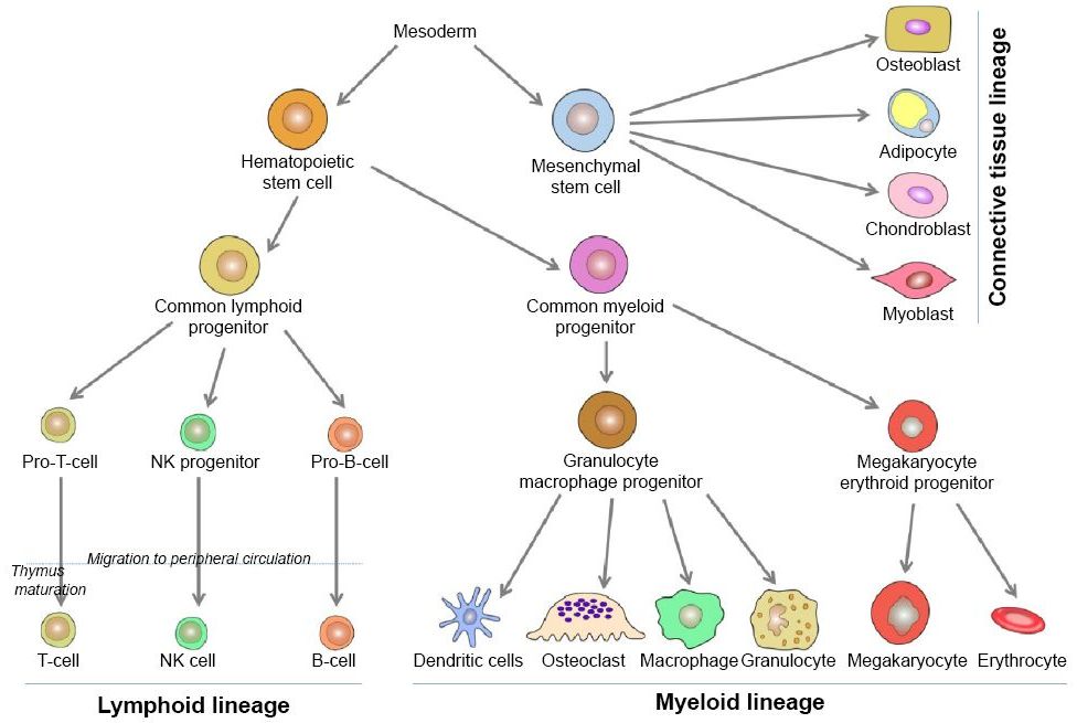
White blood cells (WBCs), also known as leukocytes, are one of the primary cells of the immune system which are responsible for fighting the disease-causing microorganisms or pathogens. Leukocytes are formed and stored in various organs of the body like spleen, bone marrow, thymus, etc. Therefore, these organs are known as “Lymphoid Organs.”

Leukocytes circulate in the body between the organs and nodes via blood vessels and lymphatic vessels; in this way, they are able to detect any foreign organism.
Leukocytes are mainly of two types;
- Phagocytes
- Lymphoid Cells/Lymphocytes
1. Phagocytes

Phagocytes are the cells which chew up invading organisms. These types of leukocytes circulating in the body have the ability to ingest and digest exogenous antigens like organisms, insoluble particles, and endogenous matters such as dead host cells, injured cells, cell debris, etc.
A. Phagocytosis
The process of engulfing foreign microorganisms by phagocytes is known as “Phagocytosis.” This process involves various steps:
- Chemotaxis involves the attraction of the phagocyte towards an organism eliciting immune responses in the body.
- The attachment of that antigen to the cell membrane of the phagocyte.
- The pseudopodia extend towards the surface attached antigens.
- With the fusion of pseudopodia, the formation of a membrane-bound structure called “phagosome” occurs.
- The antigen, then, enters the endocytic pathway of the phagocytes where the phagosome fuses with the lysosome and the formation of phagolysosome takes place.
- The lysosome contains digestive enzymes called lysozymes which finally digest the antigen.
- The digested material is then thrown out of the phagocytes via exocytosis.

B. Types Of Phagocytes
Phagocytic cells can further be classified into various types based on their cellular morphology and cytoplasmic staining characteristics.
a. Mononuclear Phagocytes
The mononuclear phagocytes consist of two types of cells, monocytes and macrophages.
i. Monocytes
During the process of “haematopoiesis” (formation of blood cells) in bone marrow, the granulocyte-monocyte progenitor cells differentiate into pro-monocyte. These pro-monocytes, then, leave the bone marrow and enter the bloodstream. There, they differentiate into mature monocytes. These mature monocytes circulate in the blood for the first 8 hours of their life, and, then, they leave the blood system, and enter various tissues of the body.
ii. Macrophages
Macrophages are the mature monocytes which settle in the tissues of the body. We can simply say that phagocytes present in the bloodstream are called monocytes while phagocytes present in the tissues are called macrophages.

When a monocyte transforms into a macrophage, its size increases five to ten times, the cell organelles gain number as well as complexity and it gains the phagocytic ability.
Macrophages disperse in the body and get settled in various organs and tissues of the system. Based upon their tissue location, macrophages are categorized as;

As described earlier, macrophages act as “Antigen Presenting Cells” (APC) but they need to be activated before they start presenting antigens on their surface.
b. Granulocytic Cells
These are those phagocytes which have a lobed nucleus as well as granular cytoplasm. They’re classified as neutrophils, eosinophils, and basophils.

i. Neutrophils

Neutrophils have a multi-lobed nucleus and their cytoplasm is granulated. They stain with both acid and basic dyes. Neutrophils are, therefore, also known as “Polymorphonuclear Leukocytes” (PMN) since they have a multi-lobed nucleus. These cells are also formed by haematopoiesis in the bone marrow. They are first released into the blood. After circulating in the blood for seven to eight hours, they finally get transferred to the tissue system. Neutrophils are the first cells to arrive at the site of inflammation. The transient increase in the number of neutrophils, called “leukocytosis,” acts as an indication of any infection in the human body. Neutrophils employ both oxygen-dependent and oxygen-independent pathways to produce antimicrobial substances. Neutrophils are more potent in digesting the microorganisms than macrophages and are able to generate a larger respiratory burst. Neutrophils contribute 50-70 percent of the total white blood cells.
ii. Eosinophils

These cells consist of a bi-lobed nucleus and granulated cytoplasm which stains with eosin red dye. Eosinophils contribute to only 1-2 percent of the leukocytes. Just like neutrophils, they also migrate from blood to the tissue space. Eosinophils play a very little role as phagocytes in engulfing the microorganisms but they play a crucial role during parasitic invasions. The substance secreted by eosinophils can destroy the cell membrane of parasites.
iii. Basophils

Basophils are the cells having a lobed nucleus and granulated cytoplasm that can be stained by methylene blue only, which is a basic. These cells constitute less than 1 percent of the total white blood cell population. Basophils are non-phagocytic cells, but are actively involved in allergic reactions. The pharmacologically active substances released by the granulated cytoplasm play an important role in certain allergic responses.
c. Mast cells
Mast cells are also involved in allergic responses. These cells, when formed in the bone marrow by haematopoiesis, start circulating in the bloodstream as a precursor of the mast cells. They get matured/differentiated once they enter the tissue system. Mast cells are present in various tissues like skin, connective tissues of various organs, and epithelial lining of the respiratory, genitourinary, and digestive tracts. Just like basophils, they also contain granulated cytoplasm with pharmacologically active substances like histamine. Mast cells and basophils together constitute the defence force of the human immune system against the allergic reactants invading the body.

d. Dendritic cells
Dendritic cells are covered with a long membranous extension which makes it appear like a dendritic cell of the nervous system, hence, its name. There are four types of dendritic cells present in our body;
- Langerhans Cells
- Interstitial Dendritic Cells
- Myeloid Cells
- Lymphoid Cells

Despite their differences, all the four types of dendritic cells express class II MHC molecules and members of the B7 co-stimulatory family.

The immature dendritic cells, also referred to as “veiled cells,” acquire antigen by the process of phagocytosis but mature dendritic cells acquire antigens by presenting those antigens on their surfaces. All four types of dendritic cells act as antigen-presenting cells (APC) and present antigen to the helper T cells (TH cells). Since they express class II MHC molecules on their surface, therefore, they’re more potent APCs than macrophages or B cells.

As soon as an antigen enters the body, the Langerhans cells and interstitial dendritic cells migrate to the lymph nodes where they express the antigen to the TH cells. This particular process is instrumental in invoking an immune response.
Another type of dendritic cell is follicular dendritic cell. These cells do not express class II MHC on their surface and, hence, do not act as antigen-presenting cells for the TH cells. They have a different role than the other four dendritic cells. They are not produced from bone marrow. These cells are present in the lymph follicles of the lymph nodes. Although they do not have the class II MHC molecules on their surface, they have membrane receptors for antibodies which allow the antigen-antibody interaction.
2. Lymphocytes

Lymphocytes contribute 20-40 percent of the body’s total white blood cell population and 99 percent to the cells in the lymph.
Lymphocytes are one of the principal cells of the immune system. They are responsible for adaptive immunity and the immunological attributes of self versus non-self recognition, memory, diversity, and specifications. We can simply say that the lymphocytes help the body to remember the previous infectious invaders, recognize them and kill them.
Lymphocytes can be further divided into B Cells, T Cells, and Natural Killer Cells (NK Cells).
A. Natural Killer Cells

Natural killer cells (also known as NK cells, K cells or killer cells) are large granular cells and do not express any surface markers, unlike B and T cells. The NK cells play a major role in host rejection of tumours and virally infected cells, i.e., they’re known for recognizing virally infected cells and detecting and controlling early signs of cancer. NK cells are also found in the placenta and protect against diseases during pregnancy.

NK cells are known for their ability to kill tumour cells without prior activation, unlike T cells which need antigen-presenting cells. Natural killer cells also release some cytokines like IFNγ and TNFα, which help in enhancing the immune responses of other immune cells like dendritic cells or macrophages.
B. B Lymphocytes

B lymphocytes got their name from their site of maturation, which is the “bursa of Fabricius” in birds. The name matched with the major site of maturation in mammalian species as well, i.e., bone marrow. B cells have membrane-bound immunoglobins (antibodies) attached on the surface. Other than immunoglobins, B cells have various other molecules present on their surface like;
• CD40- It is present on the surface of B cells and interacts with the CD40 ligand present on the surface of T helper cells. It is crucial for the survival of activated B cells and their differentiation into plasma cells and memory B cells.
• B220- It is used as a marker for B cells.
• Class II MHC molecules– They are responsible for the antigen-presenting activity of the B cells.
• CR1 (CD35) and CR2 (CD21)– These are complimentary reaction product receptors.
• B7-1 (CD80) and B7-2 (CD86)– These are the molecules that interact with CD28 and CTLA-4 which are important regulatory molecules on the surface of various T cells including the Helper T cells.
• FC γ RII (CD32)– It is a receptor of the IgG immunoglobin.

Initially, when a B cell has not interacted with an antigen, it is called a naive B lymphocyte. Same is the case with T cells; a naive T lymphocyte is the one which has not come in contact with any antigen yet. Naive B and T cells are present in the G₀ phase of the cell cycle. When any microorganism invades the body, these lymphocytes attract the antigen towards the antibodies present on their surface. The interaction of antigen-antibody activates the B and T cells. These cells then start differentiating as they move from G₀ to G1 phase and then to S, G2, and M phase. The differentiation of the B and T cells results in the formation of effector cells and memory cells.
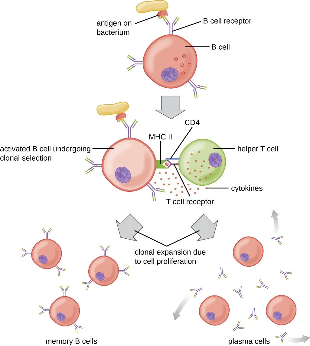
When B cells are activated, they start dividing and differentiate within 4-5 days into “Plasma Cells” and “Memory Cells.” Plasma cells have a less number of antibodies on their surface as compared to the B cells but they are responsible for synthesising and secreting antibodies against the antigens that have attacked the body. Memory cells are primarily responsible for recognizing the old antigens and storing all the pathological, morphological, and other details of those antigens in their memory in case the same antigen attacks the organism again. Plasma cells usually die within 1-2 weeks.
C. T Lymphocytes

T lymphocytes are named so because of their site of maturation which is “Thymus.” Just like the B cells, the T lymphocytes also express receptors on their surface but the T cell receptors (TCR) are structurally different from the immunoglobins. Unlike the membrane-bound antibodies present on the surface of the B cell, T cell receptors do not bind free antigens. This is the difference between humoral and cell-mediated branches of the immune system. B cells are capable of binding to the soluble antigen whereas the T cells bind only to those antigens which are present on self cells and are presented by major histocompatibility complex (MHC), tumour cells, or virally infected cells.
To be recognised by the T cells, the antigen must be present on the surface of an antigen-presenting cell via the MHC molecule. The T cells also present some membrane molecules other than antigen-binding T cell receptors, like CD3, CD4, and CD8. Other than the aforementioned, a mature T cell also presents;
• CD28- receptor for the B7 family presented on the B cell surface.
• CD45- a signal transduction molecule.
After encountering an antigen-presenting cell, T cells undergo division and differentiate into effector cells and memory cells similar to B cells. But the effector cells of the T cell lineage include cytokines secreting T helper lymphocytes and T cytotoxic lymphocytes, i.e., TH cells and TC cells.

T cells that express CD4 on their surface bind only to class II MHC molecules whereas T cells having CD8 on their surface bind to class I MHC molecules only. It also defines the functioning of the two subpopulations of T cells; CD4+ T cells generally function as T helper cells (TH) and are class II-restricted; CD8+ T cells functions as T cytotoxic cells (TC) which are restricted to the class I MHC molecules.

T helper cells, when activated, secrete cytokines that are further responsible for the activation of the B cells, T cells, and other cells involved in an immune response. This type of immune response is responsible for the production of different types of cytokines. T cytotoxic cells, when activated, start proliferation and differentiate into effector cells called cytotoxic T lymphocyte (CTL).
T suppressor cells are another type of T cells which are responsible for blocking the action of other lymphocytes so as to prevent the immune system from becoming over-active. They are also known as T regulatory cells and are being studied extensively in the treatment of cancer.

Organs of the Immune System
As discussed earlier, there are various cells, tissues, and organs which are responsible for building an immune system in an organism. Organs are the site where the cells of the immune system either mature or interact with the antigen.

Organs of the immune system are classified into two types;
1. Primary Lymphoid Organs
These are those organs where the lymphocytes mature. Once the lymphocytes are formed via haematopoiesis, they get matured in the bone marrow or thymus. The bone marrow and thymus are considered as the two primary organs of the immune system. Only when a lymphocyte has matured within a primary lymphoid organ is the cell immunocompetent (capable of producing an immune response).
T lymphocytes are matured in the thymus while B lymphocytes are matured in the bone marrow.
a. Thymus

The thymus is the site of T cell development and maturation. It is a flat and bilobed organ situated just above the heart. The thymus is externally covered with a capsule layer and internally, it is divided into lobules, which are connected to each other by a connective tissue known as “trabeculae.” Each lobule is divided into two compartments. The outer compartment or cortex is densely packed with immature T cells known as thymocytes, and the inner compartment or medulla is continuous with the network of thymocytes in the cortex.

The role of the thymus can be studied in mice by neonatal thymectomy, a procedure in which thymus is surgically removed from newborn mice. The thymectomized mice are observed and it has been seen that there is a dramatic fall in the T lymphocyte circulation in the blood and, hence, the absence of cell-mediated immunity. Ageing is accompanied by a decline in thymic function.
b. Bone marrow

Bone marrow is responsible for the maturation and development of B lymphocytes in both humans and mice. B lymphocytes originate from the lymphoid progenitor cells; proliferate and differentiate in the bone marrow.
Not all species have bone marrow as their primary lymphoid organ for B cell maturation.

2. Secondary Lymphoid Organs
Secondary lymphoid organs can be considered as the filters which monitor the content of the extracellular fluids. The fluids which are monitored are blood, lymph, and tissue fluid. These organs, then, trap any antigen present in the fluid so that the lymphocytes can interact with it. The main purpose of the secondary lymphoid organs is to provide a site to the mature lymphocytes to interact with the antigens. It is the site where lymphocytes are activated.
The secondary lymphoid organs include lymph nodes, spleen, and various mucosa-associated lymphoid tissues (MALT) such as gut-associated lymphoid tissue (GALT).
a. Lymph nodes

These are bean-shaped structures containing a large number of lymphocytes, macrophages, and dendritic cells. Lymph nodes are clustered at the junction of the lymphatic tissue. Whenever an antigen enters a tissue system, lymph nodes are the first to encounter it.
b. Spleen
The spleen plays a major role in eliciting an immune response against the antigens present in the bloodstream. It is a large, oval-shaped organ present at the upper left abdominal cavity.

Unlike the lymph nodes which are specialized in trapping the antigens from the local tissue system, the spleen filters the fluid and traps the blood-borne antigens. Spleen is supplied by the splenic artery which carries the blood-borne antigens and lymphocytes to it, unlike the lymph nodes that is supplied by the lymphatic vessels.
Spleen is also covered with capsule and is divided into compartments by trabeculae. The compartments are divided into ‘red pulp’ and ‘white pulp.’ The red pulp is the site where old and defective red blood cells are destroyed and removed.

c. Mucosal associated lymphoid tissues (MALT)

Mucous membrane lines the digestive tract, respiratory, and urinogenital system of the body. This lining is one of the major sites for pathogen entry. Therefore, the mucosa-associated lymphoid tissue has an ability to protect the body from the invading microorganisms because of a large number of plasma cells (which secrete antibodies), which is even more than the number of plasma cells present in the spleen, lymph nodes, and bone marrow combined.