
An action potential is a swift change in electrical charge across a cell membrane, seen in nerve (neurons) and muscle cells. Action potentials are vital for the proper functioning of the nervous system. It begins with cell membrane depolarizaton, driven by sodium ion influx through specific channels. This prompts potassium channels to open, causing potassium ions to exit the cell, leading to membrane repolarization. The action potential moves along the cell due to successive ion channel openings and closings. Importantly, it is an ‘all-or-nothing’ event, reaching max intensity once initiated, regardless of stimulus strength. Inputs from other neurons can affect how often action potentials occur in a given neuron. Understanding action potentials help comprehend how the body senses and responds to stimuli with precision and efficiency. For instance, when a person decides to lift their hand, a series of action potentials travel along motor neurons towards the muscle fibres responsible for hand movement. The action potential is involved in simplest to complex body movements. Here are some action potential examples found in real life:
Examples
1. Muscle contraction

Action potential controls muscle movements, allowing walking, running, or performing any physical activity. The process follows certain steps which repeat rapidly during muscle contraction and relaxation. The process begins at the neuromuscular junction, where motor neurons connect with muscle fibres. Each motor neuron enables multiple muscle fibres, forming a motor unit. When the brain sends a signal to contract a specific muscle, a nerve impulse (action potential) is generated in the motor neuron. This action potential travels along the length of the motor neuron’s axon toward the neuromuscular junction. When the action potential reaches the neuromuscular junction, it triggers the release of a neurotransmitter called acetylcholine from the motor neuron’s axon terminals. Acetylcholine is released into the synaptic cleft, the tiny gap between the motor neuron and the muscle fibre, and binds to receptors on the muscle fibre’s membrane, known as the sarcolemma. This binding causes changes in the sarcolemma’s electrical charge and initiates an action potential in the muscle fibre, which quickly spreads along the cell membrane and into the muscle fibre’s interior via a network of specialized tubules called the T-tubules. This action potential in the T-tubules signals the sarcoplasmic reticulum, a specialized organelle in the muscle fibre, to release calcium ions (Ca2+) into the muscle cell’s cytoplasm. Calcium ions bind to regulatory proteins on the muscle’s contractile proteins, actin, and myosin. This binding allows these proteins to interact and generate force, leading to muscle contraction. When the action potential ends and calcium ions are actively transported back into the sarcoplasmic reticulum, the muscle relaxes, and muscle fibres return to their resting stage.
2. Withdrawing from pain

Reflexes are essential for our safety and survival as they enable us to react quickly to potential threats or harmful stimuli. Action potentials are the electrical signals that facilitate the rapid transmission of information from sensory receptors to the central nervous system and, ultimately, to motor neurons, resulting in the reflexive response. Upon touching a hot surface, the body’s response involves the generation of action potentials in sensory neurons. The hot surface is the stimulus that initiates the process. Heat energy is detected by specialized receptors in the skin called thermoreceptors. When the thermoreceptors detect the intense heat, they undergo a change in their electrical charge. This change in charge is known as depolarization. Similarly, action potentials from pain receptors signal the sensation of pain, prompting one to react. Reflexes are triggered by a specific sensory stimulus, such as a sudden touch or pain. Withdrawing from pain involves the generation of action potentials in response to a painful stimulus. The stimulus in this case is a painful event or sensation. This could be anything from a pinprick to a burn, depending on the source of the pain. Specialized nerve endings in the skin and other tissues called nociceptors detect the painful stimulus. These nociceptors are equipped to respond to harmful or potentially damaging stimuli. Depolarization occurs when sodium ions enter the thermoreceptor cells, causing the inside of the cell to become more positively charged relative to the outside. Depolarization in the thermoreceptor cells triggers the initiation of an action potential, which then travels along the sensory neuron. These neurons are long and extend from the skin to the spinal cord. The action potential moves down the neuron like a wave, transmitting the signal to the central nervous system. Interneurons may relay the signal to motor neurons, which control muscles. Motor neurons generate their own action potentials, which lead to muscle contractions. In response to the signal, the muscles contract, causing one to quickly withdraw the hand from the hot surface. This entire process is rapid and occurs within milliseconds. It is a protective mechanism that prevents us from sustaining further injury by allowing us to react swiftly to harmful stimuli.
3. Heartbeat
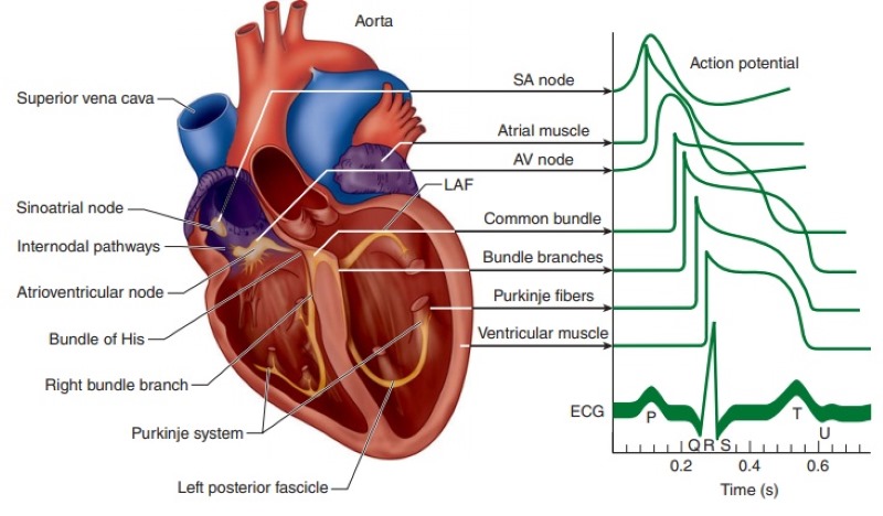
The cardiac pacemaker cells generate action potentials that regulate heart rate and rhythm. The heartbeat is initiated and regulated by specialized cells in the heart called pacemaker cells. The primary pacemaker is the sinoatrial (SA) node, located in the right atrium. Pacemaker cells generate action potentials spontaneously without external stimulation. These action potentials serve as the electrical signals that initiate each heartbeat. The action potentials generated in the SA node spread throughout the atria, causing the atria to contract and push blood into the ventricles. The electrical signals travel through specialized conduction pathways, including the atrioventricular (AV) node and the bundle of His. As the electrical signal reaches the ventricles, it causes the ventricles to contract, forcing blood out of the heart and into the circulatory system. After each contraction, the heart’s muscle cells undergo repolarization, resetting their electrical state to prepare for the next action potential. The cycle of action potentials followed by contraction and repolarization repeats rhythmically, maintaining the heart’s steady beat. The timing and coordination of action potentials in different parts of the heart are crucial for ensuring that blood is pumped efficiently and effectively throughout the body. Irregularities in the generation or conduction of action potentials can lead to heart rhythm disorders, such as arrhythmias, which can have significant health implications.
4. Digestion

Action potentials in the digestive tract muscles help move food through the digestive system. Action potentials generated by sensory receptors in the mouth and along the digestive tract play a role in the sensory perception of taste, texture, and temperature of food. These action potentials are transmitted to the brain, where they are processed, contributing to the overall eating experience. Smooth muscles in the digestive tract contract in coordinated waves (by a process known as peristalsis) to move food along the digestive tract. While action potentials are not directly responsible for initiating these contractions, they can influence and modulate the activity of smooth muscles. Action potentials from motor neurons and interneurons in the enteric nervous system (a network of nerves within the digestive tract) help regulate the timing and intensity of muscle contractions. For example, the action potentials trigger muscle contractions that mix and propel food through the digestive tract. Glands in the digestive system, such as the salivary glands, gastric glands, pancreas, and liver, release digestive enzymes, acids, and other substances in response to various signals. These signals can include action potentials triggered by sensory receptors, hormonal signals, or nervous system input. When food enters the stomach, action potentials in the stomach’s lining can stimulate the release of gastric juices to aid in digestion. Hormones such as gastrin, cholecystokinin (CCK), and secretin, which regulate digestion and nutrient absorption, are released in response to specific stimuli. Action potentials in endocrine cells can lead to the release of these hormones, which then circulate in the bloodstream and influence digestive processes in various ways.
5. Sensory perception

Action potentials in sensory neurons convey information about taste, smell, touch, sight, and hearing. Action potentials are fundamental to sensory perception as they are responsible for transmitting sensory information from sensory receptors to the brain. Specialized sensory receptors, such as photoreceptors in the eyes, mechanoreceptors in the skin, or chemoreceptors in the taste buds, are sensitive to specific sensory stimuli, such as light, pressure, or chemicals. When these sensory receptors are exposed to their respective stimuli, they undergo changes that lead to the generation of action potentials. In the case of vision, photoreceptor cells in the retina detect light and respond by generating action potentials. The sensory receptors convert the sensory input into electrical signals in the form of action potentials. This is achieved by changes in ion channels within the receptor cells’ membranes. The action potentials generated in the sensory receptors are transmitted along sensory neurons, which have long extensions (axons) that carry the electrical signals toward the central nervous system (typically the spinal cord or brain). In the central nervous system, the action potentials are processed and interpreted. This involves complex neural circuits and integration, where the brain interprets the action potentials to create our sensory experiences.
6. Breathing

Action potentials play a crucial role in the process of breathing, enabling the coordination of respiratory muscles and the regulation of breathing rate. Breathing is primarily controlled by two main groups of neurons located in the brainstem – the medulla oblongata and the pons. These neurons are part of the respiratory centres. The process of breathing begins with the generation of action potentials in specific neurons within these respiratory centres. These action potentials are spontaneous and rhythmic, initiating the breathing cycle. Once generated, the action potentials travel along nerve pathways that connect the respiratory centres to motor neurons responsible for controlling respiratory muscles. Action potentials reach motor neurons that control the contraction of respiratory muscles, which include the diaphragm and various intercostal muscles. These muscles are essential for expanding and contracting the chest cavity during breathing. During inhalation, action potentials stimulate the contraction of the diaphragm and other muscles that expand the chest cavity, allowing air to rush into the lungs. During exhalation, action potentials cease, causing these muscles to relax and the chest cavity to decrease in size, expelling air from the lungs. The rate and depth of breathing are finely tuned by the respiratory centres in response to changing oxygen and carbon dioxide levels in the blood. If oxygen levels decrease or carbon dioxide levels increase, the respiratory centres adjust the frequency and depth of action potentials to regulate breathing accordingly. While breathing is mainly an automatic process, it can also be consciously controlled to some extent. For example, one can voluntarily take a deep breath or hold it. This voluntary control involves action potentials generated by motor neurons under conscious command.
7. Swallowing

Action potentials play a role in the process of swallowing, which is a complex and coordinated action involving muscles and nerves. Swallowing is started voluntarily when one decides to eat or drink. However, once the process begins, it becomes reflexive and largely involuntary. The process starts with the sensory detection of food or liquid in the mouth and throat. Specialized sensory receptors, including mechanoreceptors and chemoreceptors, detect the presence of the material to be swallowed. When these sensory receptors detect the stimulus (e.g., food or liquid), they generate action potentials. These action potentials are transmitted along sensory neurons to the brainstem, where the swallowing reflex is controlled. In the brainstem, the sensory information from the action potentials triggers the swallowing reflex. This reflex coordinates the sequential contraction and relaxation of various muscle groups involved in swallowing. The brainstem sends action potentials along motor neurons to the muscles involved in swallowing. These muscles include those responsible for moving food or liquid from the mouth to the oesophagus (the pharyngeal muscles) and finally to the stomach (the oesophagal muscles). Action potentials in motor neurons stimulate the contraction of the relevant muscles. In the case of the pharyngeal phase of swallowing, these muscles contract to propel the food or liquid into the oesophagus while preventing it from entering the airway. In the oesophagal phase, action potentials ensure the coordinated contraction of the oesophagal muscles to move the material toward the stomach. Action potentials in the oesophagal muscles initiate a wave-like contraction pattern known as peristalsis. This pattern helps push the food or liquid down the oesophagus and into the stomach. Following the swallowing process, action potentials cease, causing the muscles involved in swallowing to relax, and the digestive process continues in the stomach.
8. Bladder control

Nerve impulses control the contraction of bladder muscles during urination. Action potentials are very important in bladder control, specifically in the coordination of muscle contractions that enable both the storage of urine and its expulsion. Specialized sensory receptors in the bladder wall, known as stretch receptors or mechanoreceptors, detect the amount of urine present in the bladder. These receptors respond to the stretching of the bladder wall as it fills with urine. When the bladder begins to fill with urine, the stretch receptors generate action potentials in response to the increased tension and stretching of the bladder wall. The action potentials generated by the stretch receptors are transmitted along sensory neurons to the spinal cord and, ultimately, to the brain. These action potentials convey information about bladder fullness and pressure. In the brain, action potentials are processed in regions responsible for conscious awareness of bladder fullness and the decision to initiate urination. These brain centres, including the pontine micturition centre, send signals back to the spinal cord to influence bladder control. The brain signals lead to the generation of action potentials in motor neurons within the spinal cord. These motor neurons control the muscles of the bladder and the urinary sphincters. Action potentials in motor neurons stimulate the contraction of the detrusor muscle, the muscle layer in the bladder wall. This contraction allows the bladder to expel urine by increasing the pressure within it. Concurrently, action potentials inhibit the motor neurons that control the urinary sphincters, which are muscles that normally keep the bladder outlet closed. This relaxation of the sphincters allows urine to flow out of the bladder and into the urethra. In most situations, the conscious decision to urinate involves action potentials generated in the brain’s control centres. When in the need to empty the bladder, action potentials are sent to the spinal cord to facilitate coordinated bladder contraction and sphincter relaxation. In infants and individuals with certain neurological conditions, action potentials can trigger reflexive bladder emptying without conscious control, known as a micturition reflex. In this case, action potentials originating from stretch receptors directly stimulate bladder contraction and sphincter relaxation when the bladder is full.
9. Coughing

When something irritates the lining of the airways, such as dust, smoke, or mucus, sensory receptors in the airways called cough receptors (or irritant receptors) are activated. These receptors detect the irritants. The activation of cough receptors leads to the generation of action potentials in sensory neurons. These action potentials transmit information about the irritation to the brainstem, specifically the medulla oblongata. In the medulla oblongata, the brainstem’s cough centre processes the sensory input and triggers a cough reflex. The cough centre generates action potentials that initiate the reflexive cough response. Action potentials from the cough centre are sent to motor neurons that control the muscles involved in coughing. These muscles include the diaphragm, intercostal muscles, and abdominal muscles. Action potentials stimulate the coordinated contraction of these muscles, which leads to increased pressure in the chest and a forceful expulsion of air. The forceful expiration of air during coughing helps expel irritants, mucus, or foreign particles from the airways, clearing the respiratory tract and protecting the lungs.
10. Sneezing

In a manner similar to coughing, sneezing is initiated by the detection of irritants, usually in the nasal passages. Sensory receptors in the nasal mucosa detect these irritants, such as dust, allergens, or pathogens. When irritants are detected, action potentials are generated in sensory neurons within the nasal mucosa. These action potentials carry the sensory information to the brainstem. In the medulla oblongata, the brainstem’s sneeze centre processes the sensory input and initiates the sneeze reflex. Action potentials generated in the sneeze centre trigger the reflexive sneeze response. The sneeze reflex involves action potentials stimulating the contraction of various muscle groups, including those in the diaphragm, chest, and throat. These muscles coordinate to produce a rapid and forceful expiration of air through the mouth and nose. The sudden and powerful expulsion of air during sneezing helps clear irritants or foreign particles from the nasal passages and respiratory tract, protecting the airways and lungs.
11. Fetal Movement

Action potentials play a role in fetal movement, allowing the developing fetus to move within the womb. The fetal movement begins when the muscles and neuromuscular connections in the developing body of the fetus reach a sufficient level of development. As the fetal nervous system matures, action potentials become capable of initiating muscle contractions. These action potentials are generated in the developing motor neurons within the fetal spinal cord and brain. Various stimuli within the womb can initiate fetal movement. This includes sensory input, such as touch or pressure, as well as internal cues related to the fetal development and changing conditions. When a stimulus triggers a response, action potentials are generated in the motor neurons that innervate the developing muscles. These action potentials travel along nerve pathways to reach the target muscles. The action potentials arriving at the developing muscles stimulate contractions. Fetal movement occurs when these muscles contract and move specific parts of the fetus, such as limbs, fingers, or toes. Fetal movement is diverse and includes actions like kicking, stretching, and general movement of limbs and body. These movements are a sign of a healthy developing nervous and musculoskeletal system. The fetal movement also helps the developing neuromuscular system strengthen and refine its connections and prepares the fetus for the motor skills it will need after birth. Action potentials in fetal motor neurons stimulate muscle contractions, allowing the developing fetus to move within the womb. These movements play a crucial role in fetal development and are a sign of the nervous system’s maturation.
12. Blinking
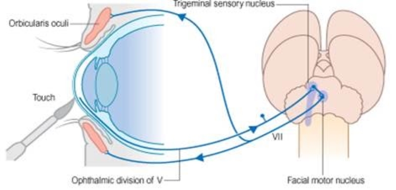
Nerve impulses cause rapid eyelid closure to protect the eye from foreign objects. The act of blinking is often initiated by the detection of a potentially harmful stimulus or change in the environment. For example, foreign particles, sudden bursts of light, or a gust of wind can trigger the blinking reflex. Specialized sensory receptors, such as corneal or ocular surface receptors, detect these stimuli. These receptors are sensitive to touch, temperature, or chemical changes on the surface of the eye. When a stimulus is detected, action potentials are generated in the sensory neurons associated with the eye’s surface. These action potentials carry the sensory information to the brain. The action potentials travel to the brain, where they are processed in regions responsible for reflexes. In the case of blinking, the brainstem is involved in the processing of sensory input related to eye protection. When the brainstem processes the sensory input and identifies it as a potential threat to the eye, it generates action potentials that initiate the blinking reflex. Action potentials generated by the brainstem stimulate motor neurons that control the muscles responsible for blinking. These muscles include the orbicularis oculi muscles, which encircle the eye. Action potentials in these motor neurons lead to the contraction of these muscles, closing the eyelids to protect the eye from potential harm. The coordinated action of these muscles results in a quick and reflexive blinking response. The duration and frequency of blinking can vary depending on the nature and intensity of the stimulus. While blinking itself is not a consciously initiated action and doesn’t involve voluntary control through action potentials, it’s a protective reflex that relies on action potentials to quickly and automatically safeguard the eyes from potential threats or irritants.
13. Sensory Adaptation

Action potentials play a role in the process of sensory adaptation, a mechanism by which sensory receptors adjust their sensitivity to sustained or repetitive stimuli. Sensory adaptation allows us to focus on new or changing stimuli while filtering out constant background information. Sensory adaptation begins when a sensory receptor detects a sensory stimulus, such as touch, odour, sound, or light. Each type of receptor is specialized to respond to specific types of stimuli. When a sensory stimulus is first detected, the sensory receptor generates action potentials in response to the change in stimulus intensity. These action potentials are transmitted along sensory neurons to the central nervous system for processing. In the case of a sustained or repetitive stimulus, the sensory receptor continues to generate action potentials as long as the stimulus is present. With time, while the stimulus is still present, the frequency of action potentials generated by the sensory receptor gradually decreases. This is a feature of sensory adaptation. During the adaptation phase, the sensory receptor sends fewer action potentials to the central nervous system in response to the constant stimulus. This decrease in action potential frequency indicates that the receptor is becoming less sensitive to the unchanging stimulus. The decrease in action potentials allows the central nervous system to filter out the constant stimulus and focus on other more important sensory information. This filtering is crucial for efficiently processing the sensory input we receive from our environment. If the stimulus intensity changes, such as an increase in pressure, brightness, or loudness, the sensory receptor quickly responds by increasing the frequency of action potentials. This alerts the central nervous system to the change in the sensory stimulus, ensuring that important new information is not missed. Sensory adaptation helps us efficiently process sensory information by reducing our response to constant stimuli, allowing us to focus on new or changing sensory input. Action potentials in sensory neurons play a critical role in transmitting these signals to the central nervous system and participating in the adaptive response to sensory stimuli.
14. Temperature Regulation

Nerve signals help regulate body temperature by prompting responses like shivering when cold. Action potentials are indirectly involved in temperature regulation through their influence on thermoregulatory responses. Temperature regulation is primarily controlled by the hypothalamus in the brain, and while action potentials themselves do not regulate temperature, they play a role in transmitting signals that initiate various temperature-related responses. The body’s temperature receptors, known as thermoreceptors, are located in the skin throughout our body. These thermoreceptors are sensitive to changes in temperature and help detect whether the body is too hot or too cold. When thermoreceptors detect a change in temperature, they generate action potentials. These action potentials are transmitted along sensory neurons to the central nervous system, primarily the hypothalamus. The hypothalamus serves as the body’s thermostat and receives sensory information in the form of action potentials from thermoreceptors. It processes this information and initiates appropriate responses to regulate body temperature. Based on the information from action potentials, the hypothalamus can trigger various effector responses to adjust body temperature. These responses include:
- Sweating: If the body is too hot, the hypothalamus stimulates sweat glands to produce sweat, which cools the skin as it evaporates.
- Shivering: If the body is too cold, the hypothalamus activates muscles to shiver, generating heat through muscular contractions.
- Vasodilation and Vasoconstriction: The hypothalamus can also adjust blood vessel diameter, increasing or decreasing blood flow to the skin, to either release or conserve heat, respectively.
As the effector responses are initiated, the hypothalamus continuously monitors the body’s temperature through ongoing input from thermoreceptors. If the body’s temperature approaches the desired set point, the hypothalamus adjusts the effector responses accordingly. While action potentials themselves do not directly control temperature, they are instrumental in relaying temperature-related sensory information from the thermoreceptors to the hypothalamus. The hypothalamus processes this information and triggers the appropriate physiological responses to maintain the body’s temperature within a narrow range, ensuring homeostasis.
15. Pupil Constriction

Action potentials are involved in the process of pupil constriction, a crucial mechanism of the visual system that regulates the amount of light entering the eye. The size of the pupil is controlled by the autonomic nervous system, which relies on action potentials to transmit signals. The amount of light entering the eye is detected by specialized cells in the retina called photoreceptors, specifically the rod and cone cells. When light intensity increases, photoreceptors generate action potentials that carry visual information to other neurons in the retina. These action potentials are transmitted along the optic nerve to the brain. In the brain, the visual information is processed in the visual cortex and other regions responsible for visual perception and reflexes. The brain processes the information and determines the appropriate response based on the level of illumination. The brain relays signals to the autonomic nervous system to initiate the pupillary light reflex. The autonomic nervous system consists of two branches: the sympathetic and parasympathetic systems. Pupil constriction, or miosis, is primarily controlled by the parasympathetic nervous system. Action potentials generated in parasympathetic neurons are transmitted to the circular muscle of the iris, which encircles the pupil. The parasympathetic neurons release the neurotransmitter acetylcholine, which binds to receptors on the circular muscle cells. When acetylcholine binds to its receptors, it triggers action potentials in the circular muscle cells, leading to their contraction. This causes the pupil to constrict, reducing its size and decreasing the amount of light entering the eye. The reduction in pupil size due to constriction helps protect the retina from excessive light exposure, preventing potential damage in brightly lit environments. The pupillary light reflex is an adaptive mechanism that allows the eye to quickly adjust to changing light conditions, ensuring optimal vision and protecting the sensitive retinal cells.
16. Gag Reflex

The gag reflex, also known as the pharyngeal reflex, is a protective mechanism that involves action potentials and helps prevent foreign objects from entering the throat and airway. The gag reflex is typically triggered by the presence of an irritant or foreign object at the back of the throat or in the oral cavity. This could be anything that may pose a threat, such as a piece of food, liquid, or any other object. Specialized sensory receptors located in the back of the throat, on the soft palate, and in the uvula (the dangling structure at the back of the throat) detect the presence of the irritant. These sensory receptors are capable of detecting touch and pressure. When the sensory receptors detect a foreign object or irritant, they generate action potentials in sensory neurons. These action potentials transmit the sensory information to the brainstem. The action potentials travel to the brainstem, specifically, the medulla oblongata, which is responsible for many involuntary reflexes, including the gag reflex. In the brainstem, the sensory information is processed, and action potentials are generated to initiate the gag reflex. If the brainstem interprets the sensory input as a potential threat, it sends action potentials to trigger the reflexive response. Action potentials in motor neurons control the contraction of muscles involved in the gag reflex. The muscles that contract include those in the pharynx, soft palate, and throat. These contractions work to push the irritant away from the sensitive structures at the back of the throat and toward the mouth. The coordinated muscle contractions generate the gagging response, which typically involves retching, coughing, or the expulsion of the irritant from the throat and into the mouth. The gag reflex serves a protective function by preventing foreign objects or irritants from entering the airway and potentially causing choking or aspiration (inhalation of foreign material into the lungs). The gag reflex is a rapid and automatic response initiated by action potentials in response to sensory input. It helps safeguard the airway and expels potentially harmful or irritating substances from the throat.
17. Balance and Equilibrium

Action potentials from inner ear hair cells contribute to balance and spatial orientation. Action potentials are not directly involved in the sense of balance and equilibrium but rather play a role in transmitting sensory information from balance-related structures in the inner ear to the brain. The sense of balance and equilibrium is primarily controlled by the vestibular system, which consists of fluid-filled structures and hair cells in the inner ear. Within the inner ear, there are specialized sensory receptors known as hair cells. These hair cells are part of the vestibular system and are sensitive to changes in head position and motion. When the head or body is moved, the movement of fluid within the inner ear stimulates the hair cells. This stimulation activates the hair cells to generate electrical signals, including action potentials. The action potentials generated by the hair cells are transmitted along sensory neurons in the vestibular nerve. This nerve carries the electrical signals to the brainstem. In the brainstem, the sensory information from the vestibular nerve is processed to determine the head’s position, movement, and orientation in space. The brainstem, specifically the vestibular nuclei, plays a crucial role in this processing. The processed information from the vestibular system is integrated with visual input and proprioceptive information (sensory information from muscles and joints). This integration allows the brain to create a comprehensive perception of balance and spatial orientation. In addition to reflexive actions, the brain uses the sensory information from the vestibular system to provide a conscious sense of the body’s position and movement in space.