
In microbiology, bacteriophages hold immense value and application. Often referred to simply as phages, these microscopic entities represent a unique group of viruses that specifically target and infect bacteria, playing a vital role in shaping bacterial populations and ecosystems. Bacteriophages come in a diverse array of shapes, sizes, and genetic compositions. Based on their morphology, bacteriophages can be broadly categorized into tailed phages (characterized by a distinctive icosahedral head connected to a long tail) and non-tailed phages (various shape ranges, including spherical, filamentous, pleomorphic, etc.). Within these broad categories, bacteriophages are further classified into various families and genera based on their genetic material, life cycles, and other structural features. Lytic (T2, T4, T7, P1, and PhiX174 phages) and lysogenic bacteriophages (Lambda, P22, P1, Mu, and PhiC31 phages) are two different life cycle strategies employed by bacteriophages to infect and reproduce within their bacterial hosts. Lytic bacteriophages are often called ‘virulent’ phages because after taking over their host’s machinery, the infection cycle ultimately leads to the death (lysis) of the host bacterial cell. Lysogenic bacteriophages are known as temperate phages and utilize a more complex life cycle that involves integrating their genetic material into the host bacterium’s genome. The host cell reproduces along with the integrated phage DNA (prophage), which can exit the host genome under certain conditions to initiate the lytic cycle. Lysogenic bacteriophages provide an advantage to the host bacterium by extending new genetic traits to the bacterium. This simultaneously leads to an increased chance of survival and virulence for the phage. However, the transition from the lysogenic to the lytic cycle is essential for the release of phage particles and the continuation of the phage cycle. Based on these types, various bacteriophage examples are found in nature that exhibit diverse strategies for infecting and manipulating bacterial hosts.
Examples
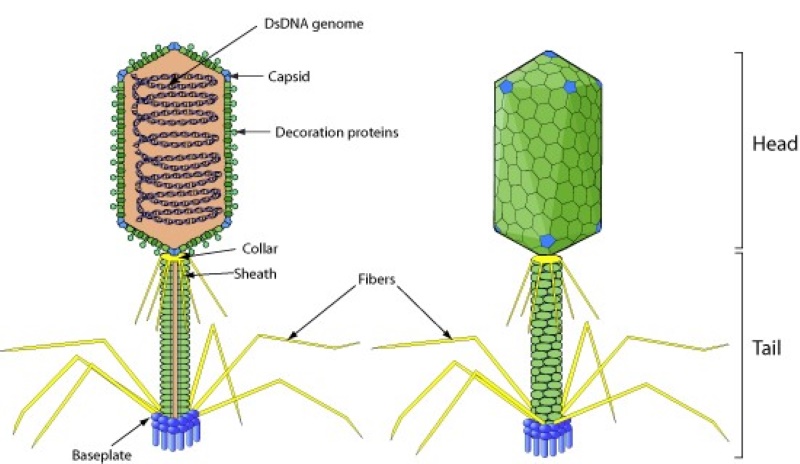
1. T4 Bacteriophage

The T4 bacteriophage, also known as Enterobacteria phage T4, is a member of the family Myoviridae (tailed bacteriophages). These viruses have a distinct appearance with an icosahedral head and a long contractile tail. T4 is renowned for its role as a model organism in the study of bacteriophages and the molecular mechanisms underlying their interactions with bacterial hosts. T4 bacteriophages naturally inhabit environments where their host bacterium, Escherichia coli (E. coli), is present (in the intestines of humans and animals). T4 phages have a complex structure. Their head contains the genetic material, which is a linear double-stranded DNA molecule, and their long tail aids in attachment to the host cell. T4 bacteriophages use specialized tail fibers to attach to specific receptors on the E. coli cell surface, followed by the injection of their genetic material into the host cell. Once inside the host cell, T4 takes control of the bacterial machinery, directing it to replicate the phage DNA and produce new phage particles. The T4 replication cycle is highly regulated, with precise timing and coordination of events. T4 follows a lytic lifecycle, meaning that it ultimately leads to the lysis (bursting) of the host E. coli cell, releasing a multitude of newly formed T4 phage particles. T4 has been employed as a tool in molecular biology and genetic engineering. Its DNA polymerase, ligase, and other enzymes have been used in various molecular techniques. T4 phages are used as vectors for cloning and expressing foreign genes in bacteria. They serve as vehicles for introducing specific genes into bacterial hosts for research and industrial applications. T4’s well-characterized genetics and genome have made it a favoured subject for studying gene regulation, mutagenesis, and genetic recombination. T4 and related bacteriophages are being investigated for their potential use in phage therapy (a promising alternative to antibiotics). Researchers are exploring their effectiveness in treating bacterial infections, including antibiotic-resistant strains.
2. Lambda Phage

The Lambda phage (formally known as Bacteriophage lambda or Lambda (λ)) is a member of the Siphoviridae family of tailed bacteriophages. Lambda phage can seamlessly transition between lytic and lysogenic cycles, making it a captivating subject of study in microbiology and molecular biology. Lambda phages are commonly found in the intestinal tracts of humans and animals, along with its host, E. coli. Lambda phages possess an icosahedral head containing their genetic material, which is a linear double-stranded DNA molecule. A long, non-contractile tail with tail fibers is connected to the head that facilitates attachment to specific receptors on the E. coli cell surface. Lambda phage has the ability to follow a bifurcated life cycle. Upon infecting an E. coli cell, Lambda can choose between the lytic cycle, where it replicates and causes host cell lysis, or the lysogenic cycle, in which it integrates its DNA into the host genome. This allows them to become a prophage and coexist with the host without immediate destruction. Lambda phage serves as an invaluable model organism in molecular biology and genetics. Its capacity to switch between lytic and lysogenic states is highly significant in studying gene regulation, DNA recombination, and genetic switches. The lambda switch is a classic example of how a phage can toggle between two different developmental pathways based on environmental cues. Lambda phage offers a significant role in fundamental research, giving insights into virus-host interactions, gene regulation, and the dynamic balance between lytic and lysogenic states in the microbial world. Lambda phages have also been utilized as a tool in genetic engineering where they are modified as vectors to introduce foreign DNA into host bacteria for various experimental and biotechnological purposes, including the creation of recombinant DNA molecules. Lambda phage and related temperate phages have been explored in phage therapy, aiming to combat bacterial infections, including antibiotic-resistant strains. Understanding their lysogenic cycles can provide insights into the potential use of temperate phages as therapeutic agents.
3. T7 Bacteriophage

The T7 bacteriophage (Enterobacteria phage T7) is a captivating example of viral specialization in the microbial world. It belongs to the Podoviridae family (tailed bacteriophages). T7 bacteriophages inhabit the intestines of humans and animals, a natural habitat of its bacterial host, E.coli. T7 phages exhibit a distinctive structural design. They boast an icosahedral head housing their genetic material, which is a linear double-stranded DNA molecule. Unlike some phages with long tails, T7 has a relatively short tail (a distinctive characteristic of the Podoviridae family). T7 utilizes its specific tail fibers to attach to precise receptors on the E. coli cell surface, followed by the injection of its genetic material into the host cell. T7 bacteriophage’s well-characterized genetics and rapid lytic cycle make it a valuable tool for studying fundamental molecular processes, such as DNA replication, transcription, and translation. T7 RNA polymerase, an enzyme encoded by T7 phage genes, is used extensively in molecular biology laboratories. It allows researchers to synthesize RNA from DNA templates, making it a crucial component in in-vitro protein expression systems. Modified T7 phages are harnessed as vectors in genetic engineering. T7 RNA polymerase-based systems have been instrumental in the development of various biotechnological applications, including the production of recombinant proteins, RNA synthesis, and the study of gene expression regulation. T7 bacteriophages are used for phage display techniques to select specific proteins with affinity for particular targets. This has implications for drug development and diagnostics.
4. M13 Bacteriophage

The M13 bacteriophage or M13 phage, is a small filamentous bacteriophage that belongs to the Inoviridae family (filamentous bacteriophages). It is known for its simplicity, ease of manipulation, and utility in a wide range of genetic and molecular studies. M13 bacteriophages are found naturally in the human intestinal tract, along with their bacterial host, E. coli. M13 phages have a long thin filamentous body, typically about 900 nanometers in length, with a single-stranded DNA genome enclosed within a protective coat protein. M13 phage plays a significant role as a cloning vector in molecular biology and in DNA sequencing techniques. The single-stranded DNA of M13 can be sequenced directly, and it has been used in the Sanger sequencing method, a classic DNA sequencing approach. M13 is widely used in phage display technology, allowing the presentation of foreign proteins or peptides on its surface. Phage display is crucial in drug discovery and the study of protein-protein interactions. M13 also serves as a model for studying DNA replication, recombination, and gene regulation. Researchers have used M13 phage to study bacterial evolution and adaptation in response to changing environmental conditions. By subjecting E. coli cultures to M13 infection, scientists can investigate how bacteria evolve resistance to phage infection.
5. P1 Phage

The P1 bacteriophage (P1 phage) is a specialized bacteriophage with the ability to integrate its genetic material into the host bacterium’s genome (transduction). This feature, along with its ability to form lysogenic relationships with its host, Escherichia coli (E. coli), has made P1 phage an invaluable tool in bacterial genetics and molecular biology. P1 phages naturally inhabit environments where their host bacterium, E. coli, thrives, i.e., in the human intestinal tract. P1 phages belong to the family Siphoviridae and consist of an icosahedral head containing their genetic material, which is a double-stranded DNA molecule, and a long non-contractile tail that aids in attachment to the host cell. During the lysogenic cycle, P1 can incorporate a portion of the host’s DNA into its genome. When it subsequently switches to the lytic cycle, it can package this bacterial DNA and transfer it to another E. coli host cell. This process, known as transduction, has been a valuable tool for transferring specific genes or markers between bacterial strains. P1 phage-mediated transduction has been crucial in genetic mapping studies, allowing researchers to determine the relative positions of genes on a bacterial chromosome. By transferring specific markers, scientists can create genetic maps that provide insights into the organization of bacterial genomes. P1 phage-based techniques have also been used for gene knockout experiments in which the researchers can introduce a P1 phage carrying a modified or disrupted version of a specific gene into a bacterial host, effectively inactivating that gene and studying its effects on the host’s phenotype. In recombinant DNA technology, P1 phage can serve as a vector for transferring genes of interest into E. coli or other related bacteria. This allows the introduction of foreign genes into bacterial hosts for various biotechnological purposes.
6. Mu Phage

Bacteriophage Mu phage, also known as phage Mu or Mu (μ), belongs to the Myoviridae family. Unlike many other bacteriophages, Mu is not solely dedicated to infecting bacterial cells and reproducing within them, instead, it behaves as a mobile genetic element that can integrate into and excise from the host bacterium’s genome, leading to various genetic mutations and rearrangements. Like their other counterparts, Mu phages inhabit the intestines of humans and animals along with their host bacteria, E.coli. Mu phage have a typical tailed bacteriophage structure with an icosahedral head containing its genetic material, which is a double-stranded DNA molecule. The head is connected to a long tail, allowing for attachment to the host cell and DNA transfer. Mu phage is unique in its ability to undergo a process called transposition, which is the movement of genetic material (Mu DNA) within the host genome. Mu phage can integrate its DNA into the host bacterium’s chromosome and then excise from it, potentially taking nearby host genes with it during the process. The transpositional activity of Mu phage can result in significant genetic rearrangements in the host chromosome. These rearrangements include inversions, duplications, and deletions of DNA segments, which contribute to genetic diversity and evolution in bacterial populations. Mu phage has been a valuable tool for studying the process of transposition in bacteria. Its ability to move and rearrange DNA within the bacterial genome has provided insights into the molecular mechanisms underlying genetic recombination. Mu phage is also used as a tool for gene knockout experiments where Mu phage is introduced with modified or disrupted genes into a host bacterium. Due to this, specific genes can be inactivated, allowing scientists to study their function or the consequences of their absence. The transposition activity of the Mu phage displays the bacterial evolution and adaptation history. It is useful to study new genetic traits and the generation of diversity within bacterial populations.
7. T2 Bacteriophage

The T2 bacteriophage (T2 phage or Enterobacteria phage T2) is a well-studied member of the bacteriophage family. T2 bacteriophage has long served as a model organism in bacteriophage research. Its simple lytic life cycle, well-defined genetic makeup, and rapid replication make it an ideal subject for studying phage-host interactions and molecular mechanisms. E. coli, the main host bacterium for T2 bacteriophage, is a common inhabitant of the human and animal intestinal tracts, providing a natural habitat for T2 phage. T2 phages possess a tailed bacteriophage structure with an icosahedral head containing double-stranded DNA as their genetic material. The head is connected to a long tail, which facilitates the attachment to specific receptors on the E. coli cell surface. T2 phage played a crucial role in early studies of DNA replication. The pioneering experiments of Martha Chase and Alfred Hershey, known as the Hershey-Chase experiment (1952), used T2 phage to demonstrate that DNA is the genetic material of phages instead of protein. T2 phage has been used for genetic mapping studies through which its ability to recombine with the host genome allowed researchers to create genetic maps, providing insights into the organization of bacterial and phage genomes. While not as widely explored as some other phages for phage therapy, T2 and related bacteriophages have been investigated for their potential in treating bacterial infections, including antibiotic-resistant strains.
8. Vibrio Phage
Vibrio phages are a group of bacteriophages that specialize in infecting and preying upon bacteria of the Vibrio genus. There is considerable diversity among Vibrio phages in terms of their genetic makeup, morphological features, and infection mechanisms. Some belong to the families Myoviridae, Podoviridae, and Siphoviridae, exhibiting variations in their structures and lifestyles. Vibrio phages have been investigated as potential biocontrol agents for Vibrio cholerae, the pathogen responsible for cholera. Monitoring Vibrio phages can aid in disease surveillance and early detection of Vibrio-related outbreaks. Vibrio phages are found in environments where Vibrio bacteria thrive, which is often in aquatic ecosystems such as marine and estuarine environments. Vibrio bacteria are commonly associated with seawater, making it a natural habitat for Vibrio phages. Vibrio phages are essential tools for studying the dynamics of bacterial populations in aquatic ecosystems. They can influence the abundance and diversity of Vibrio bacteria, shedding light on the ecological roles of phages in shaping microbial communities. Vibrio phages have a diverse genetic makeup and serve as model organisms for studying fundamental phage biology, including DNA replication, transcription, and protein expression.
9. PhiX174

PhiX174 phage, or Enterobacteria phage PhiX174, is a small single-stranded DNA (ssDNA) bacteriophage with a distinctive icosahedral head containing its genetic material. It lacks a conventional tail structure but has unique assembly proteins that assist in the formation of mature phage particles. Despite its small size, PhiX174 has played a significant role as a model organism for studying DNA replication, DNA sequencing, and the development of DNA sequencing methods. It shares its natural habitat with its host bacteria E.coli in the intestinal tracts of humans and animals. In 1977, Frederick Sanger and his team used the dideoxy sequencing method (Sanger sequencing) to determine the complete nucleotide sequence of PhiX174, marking a groundbreaking achievement in genomics. Hence, PhiX174 phage played a crucial role in the development of DNA sequencing techniques, and its DNA is used as a control in DNA sequencing experiments. Its known sequence serves as a reference point, helping researchers validate and calibrate their sequencing instruments and procedures. PhiX174 continues to be used in molecular biology research, particularly in studies involving ssDNA viruses and the mechanisms of viral replication. Its compact genome and well-characterized biology make it a valuable subject for understanding fundamental biological processes. Moreover, PhiX174 has been used in phage display techniques, which allow the presentation of foreign peptides or proteins on its surface. This has applications in the selection of specific binding molecules, such as antibodies, peptides, or enzymes, for various research and biotechnological purposes.
10. PRD1

The PRD1 phage, also known as Enterobacteria phage PRD1, belongs to the Tectiviridae family and is known for its unique structure. It has an icosahedral protein capsid that encloses its genetic material, which is a double-stranded DNA genome. What sets PRD1 apart is that its genome is enveloped by a lipid membrane, a feature rarely found among bacteriophages and more commonly associated with cellular organisms. PRD1 phage inhabits environments where its host bacterium, typically Escherichia coli (E. coli), or other Gram-negative bacteria, is present. Such environments include soil, water, and the intestinal tracts of humans and animals. PRD1 phage has been used as a model system for studying the molecular mechanisms of virus-host interactions. Its membrane and unique genome packaging provide insights into how viruses enter and exit host cells and how they deliver their genetic material. PRD1’s lipid membrane has potential applications in biotechnology. Researchers have explored the use of PRD1 phage as a delivery vehicle for therapeutic molecules, such as drugs or genes, taking advantage of its ability to interact with cellular membranes. PRD1’s protein capsid is also of interest in structural biology. The capsid’s icosahedral symmetry and stability have made it a subject of study for understanding the principles of protein-based nanostructures.
11. Cyanophage

Cyanophages (cyanobacterial viruses) specifically infect cyanobacteria, the photosynthetic microorganisms responsible for the production of a significant portion of the Earth’s oxygen. Cyanophages play a significant role in aquatic ecosystems, where cyanobacteria are abundant. They exhibit a remarkable diversity in both genetic makeup and morphology, showcasing their adaptability to a wide range of environmental conditions. Their lifecycle typically involves the infection and lysis of cyanobacterial hosts, releasing new cyanophages into the water column. The significance of cyanophages extends beyond their role as regulators of cyanobacteria. They also have a profound impact on global biogeochemical cycles and climate regulation. Cyanobacteria, through photosynthesis, remove carbon dioxide from the atmosphere, and cyanophages influence this process by controlling their cyanobacteria populations. Additionally, the lysis of cyanobacterial cells releases organic matter and nutrients back into the water, influencing the nutrient balance and the growth of other microorganisms. Some cyanophages carry auxiliary metabolic genes (AMGs) that can affect cyanobacteria’s metabolic pathways, potentially influencing carbon and nutrient cycling on a global scale.